Journal
Same Device, Different Surgery — On the Inferior Turbinate
Nose
The equipment has become simple. Who performs the surgery, and how, still matters.
A few terms first
Inferior turbinate — a long structure attached to each side wall of the nose, running from just inside the nostril to deep at the back of the nasal cavity. It moistens, warms and filters the air you breathe in.
Inferior turbinate reduction — surgery that shrinks a swollen turbinate to widen the airway. It is often called "rhinitis surgery."
Coblator (radiofrequency) — a surgical device that reduces tissue volume using radiofrequency energy.
Empty nose syndrome — a condition in which the nose is wide open, yet airflow can no longer be felt, so breathing feels blocked and suffocating.
Just ten years ago in Korea, rhinitis surgery — and turbinate surgery in particular — was the domain of ENT (ear, nose and throat) surgeons. A few practitioners of traditional Korean medicine occasionally applied treatments such as bee-venom acupuncture to the turbinates, but ENT surgeons were the main providers.
Over the past four to five years, many doctors who perform rhinoplasty have begun reducing the inferior turbinates during rhinoplasty using a radiofrequency device called a Coblator. It is very easy to use, so any doctor who performs rhinoplasty and knows the anatomy of the nose can do it.
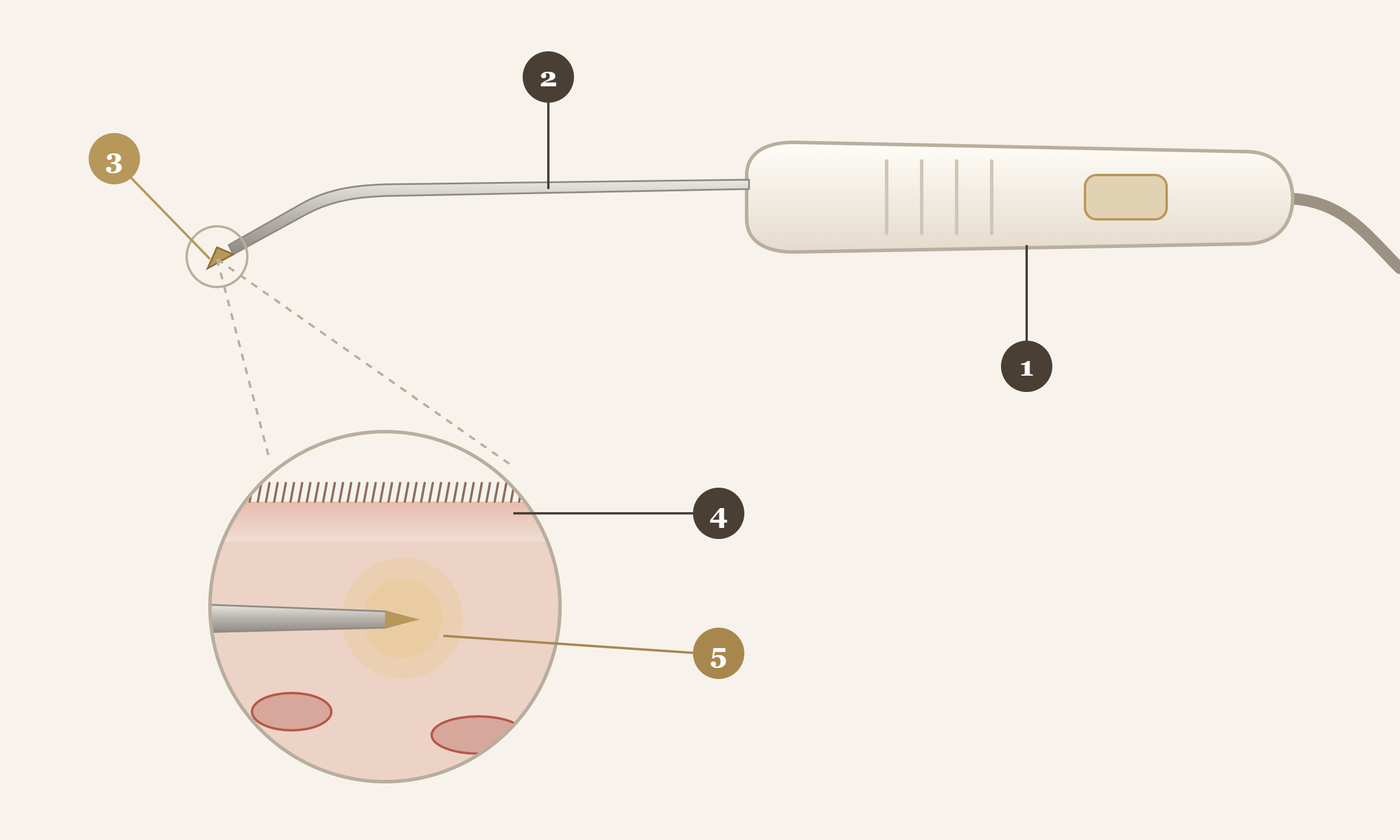
Even so, however simple the equipment becomes, you should know that a patient's quality of life after surgery can differ greatly depending on who performs it, and how.
A nose that is blocked again after six months
There is something I often hear in the consultation room: "I had rhinitis surgery along with my rhinoplasty six months ago, but my nose is blocked again."
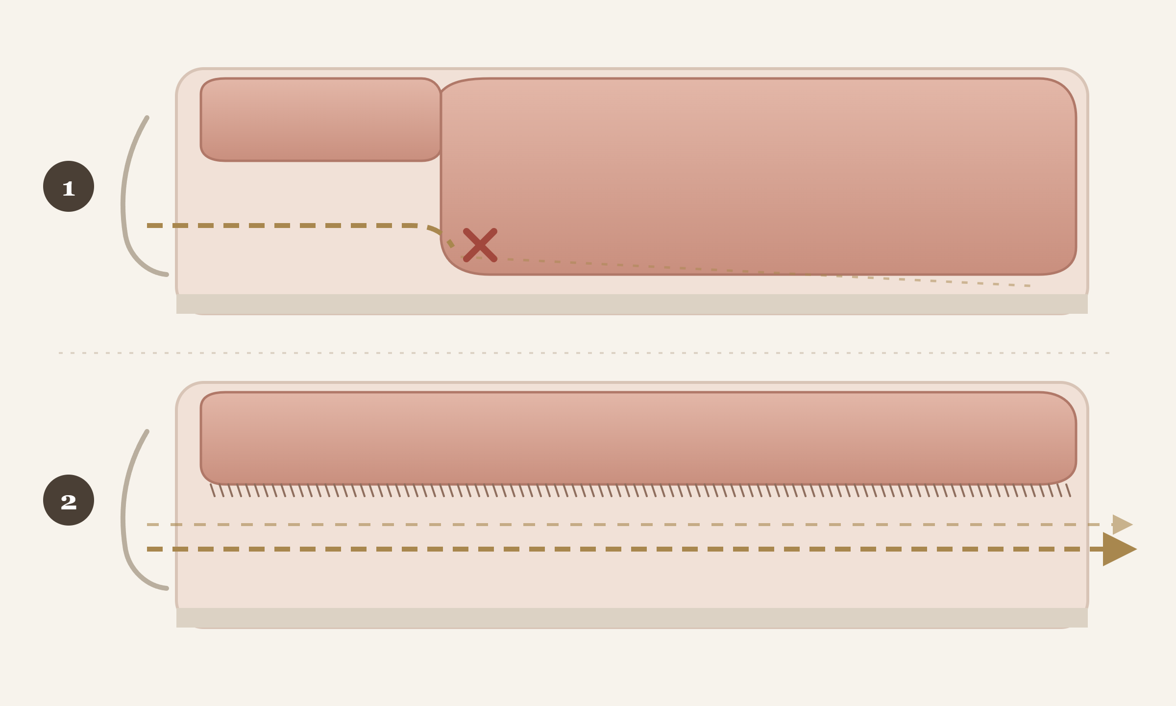
When I look at the CT and endoscopy of these patients together, I often find that only the front part of the turbinate was slightly reduced. It is less that the surgery itself went wrong, and more a difference in how the structure of the turbinate was understood and treated.
In turbinate surgery, there are three things I am always careful about.
Reduce the entire length evenly, not just the front.
Protect the cilia and receptors on the surface.
Avoid the large blood vessels deep inside.
First: reducing only the front does not open the airway
The inferior turbinate is a cylindrical structure that runs from near the nostril all the way to the back of the nose. Air flows through the nose along this entire cylinder.
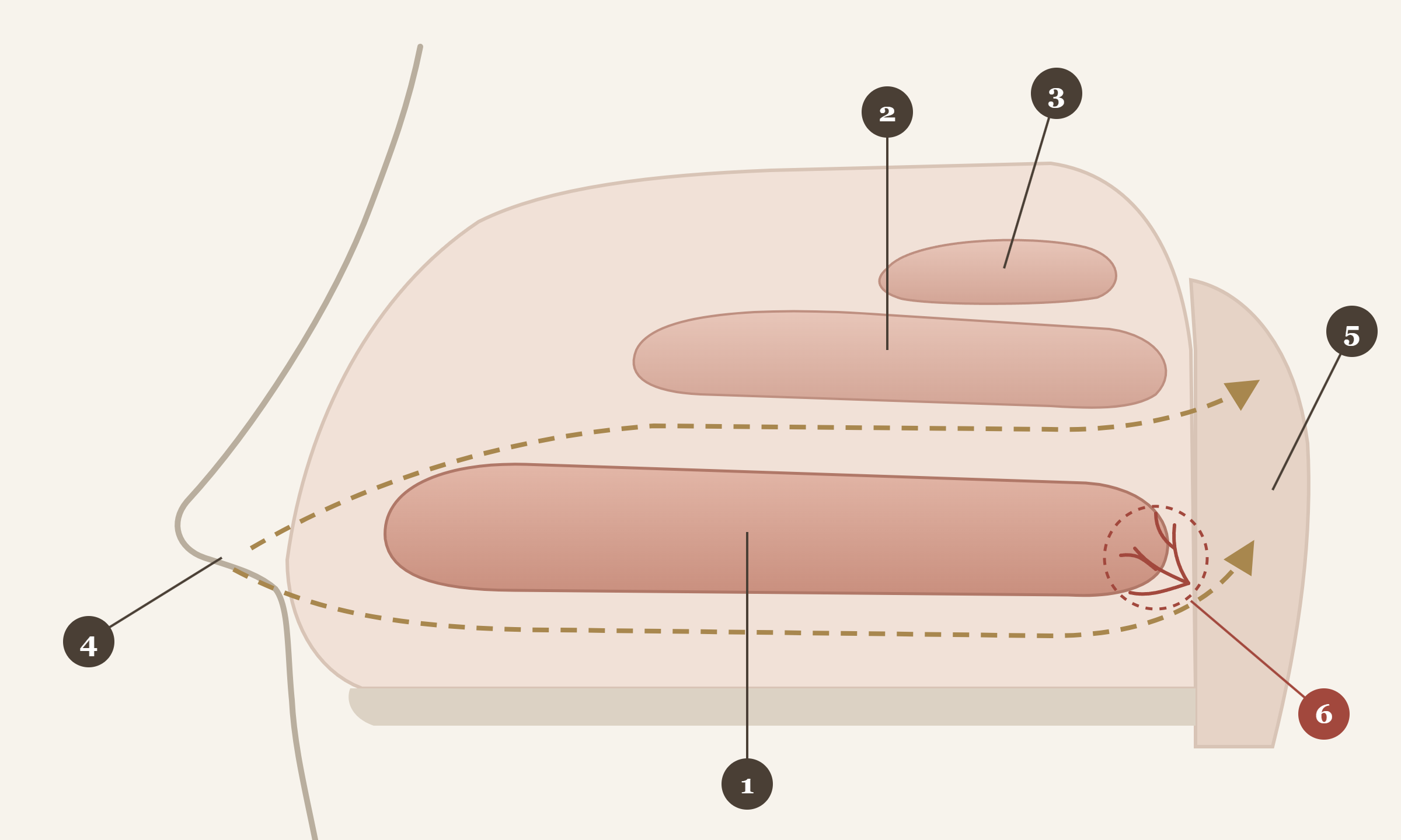
So reducing only the front end near the nostril does not fully clear the airway. It needs to be reduced evenly along its entire length — starting from the back, through the middle, to the front. It sounds simple, but working precisely deep inside the nose is harder than it seems.
Second: once the surface is damaged, it cannot be undone
Under a microscope, the surface of the turbinate is covered with countless cilia. This surface moistens, warms and filters the air you breathe in, protecting the cells of the lungs from cold, dry air.
It has one more, lesser-known function: receptors that sense air passing through. The feeling that "I can breathe" when you inhale and exhale is the signal these receptors send.
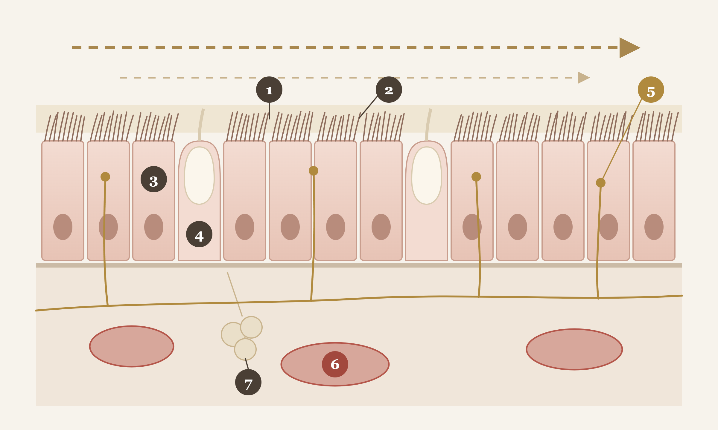
Excessive radiofrequency cauterization shrinks the turbinate quickly. But if the surface is damaged in the process, the receptors are destroyed with it. The same applies when the surface is broadly cauterized with an instrument such as bipolar forceps rather than a radiofrequency device — and in that case, far more crusting occurs.
When the receptors are destroyed, the nose is wide open, yet air passing through can no longer be felt. The brain interprets this as a blocked nose. This is called empty nose syndrome.
When the breath they have felt naturally all their lives suddenly can no longer be felt, patients describe it as suffocating, like drowning. Some describe a terror of being unable to breathe. It shakes every part of daily life.
Once destroyed, the receptors do not recover. To restore the lost volume, methods such as inserting rib cartilage, inserting it in crushed form, or injecting stem cells obtained from fat have been studied, and I took part in that research myself [1]. Even so, it remains a condition that is truly difficult to improve over the long term.
That is why protecting the surface matters above all. How strongly and how long the radiofrequency is applied, and how long it rests — each setting of power and time decides the result.
Third: large blood vessels run deep inside
I said the turbinate should be properly reduced all the way to the back, but the back of the nose is a place that calls for care. Branches of several arteries supplying the face and head pass through here, and near the rear end of the inferior turbinate is a cluster of vessels called Woodruff's area.
The tip of a radiofrequency device is not as sharp as a needle, but it is sharp enough to scrape or puncture the mucosa. If this area is injured or cauterized too deeply, heavy bleeding can occur suddenly — not right after surgery, but days or one to two weeks later, when the crusts fall off.
Bleeding from a vein usually stops when the nose is packed. Arterial bleeding is different. Blood can pour not only from the nostrils but into the mouth, and a hemoglobin level of 12–14 can drop to 6–7. It is a very dangerous situation.
If worry about such bleeding leads a surgeon to reduce only the front or middle, we are back to the first problem: the surgery is less effective.
A simple surgery, but not one to take lightly
Inferior turbinate reduction looks like a simple procedure anyone with the equipment can perform. But reducing the whole cylinder evenly, protecting the cilia and receptors on the surface, and avoiding the vessels deep inside must all be achieved together.
Only with a deep understanding of the inside and outside of the turbinate, its position and the surrounding structures can it be reduced safely — and the comfort of breathing after surgery lasts longer. Only for those who truly need it, only as much as needed, exactly where it is needed.
If you have already had surgery and are still uncomfortable, endoscopy and CT can show the current state inside your nose. If the nose is sufficiently open but the stuffiness continues, it is worth checking whether another cause, beyond the nose, is involved.
Anyone can use the equipment. What must be preserved is something the equipment will not tell you.
Dr. Soon Be Hong
Reference
[1] Kim DY, Hong HR, Choi EW, Yoon SW, Jang YJ. Efficacy and Safety of Autologous Stromal Vascular Fraction in the Treatment of Empty Nose Syndrome. Clin Exp Otorhinolaryngol. 2018;11(4):281–287. doi: 10.21053/ceo.2017.01634 · PMID: 29764011
Co-author Hong HR is Dr. Soon Be Hong, who took part in the study under her former name; the paper lists her by that name.
A prospective study at the Department of Otolaryngology, Asan Medical Center (Seoul), in which the stromal vascular fraction (SVF — a cell fraction containing stem cells) obtained from each patient's own abdominal fat was injected into both inferior turbinates of 10 patients with empty nose syndrome. Some inflammatory markers in nasal secretions decreased after injection, but a single injection did not clearly improve symptoms or satisfaction, and the authors concluded that further research into better regenerative treatments is needed.
Last updated